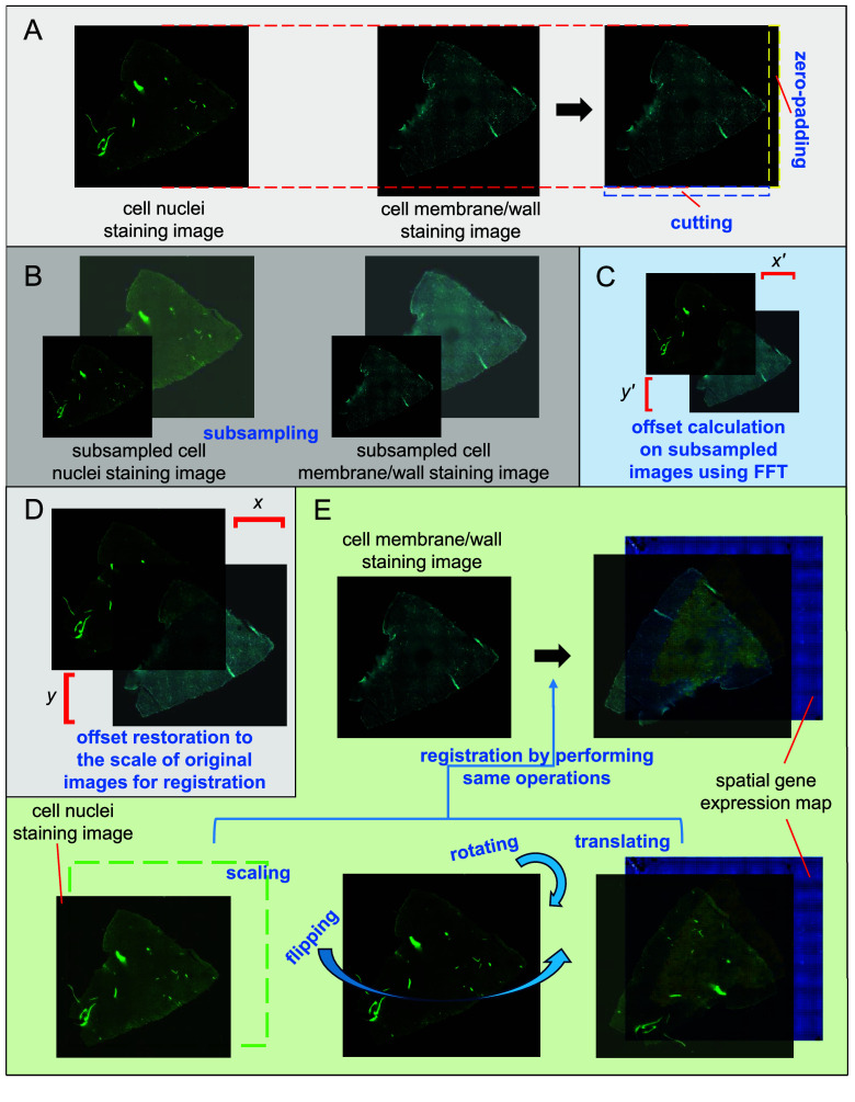
Figure 2.
Registration of the cell membrane/wall staining image and spatial gene expression map using the cell nuclei staining image as a bridge. (A) The size of the cell membrane/wall staining image is adjusted to be consistent with the cell nuclei staining image. (B) The cell nuclei and cell membrane/wall staining images are subsampled. (C) Calculation of the offsets of the subsampled images. (D) Restoring the offsets to the scale of the original images for registration. (E) Registration of the spatial gene expression map and cell nuclei staining image by performing scaling, rotating, flipping, and translating, followed by the registration of the spatial gene expression map and cell membrane/wall staining image by performing the same operations.

