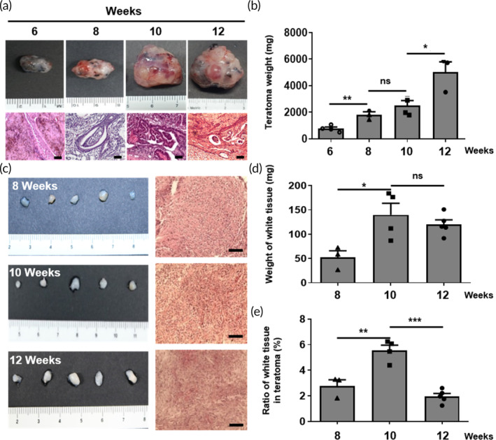
FIGURE 2.

Characterization of teratomas and condensed MSC mass from human iPSC‐derived teratomas during teratoma development. (a) Macroscopic appearance and histological examination of teratomas as a function of post‐implantation time (weeks). Scale bar, 100 μm. (b) Comparison between the weights of teratomas as a function of post‐implantation time (weeks). The data are presented as the means ± s.d. (n = 6, biologically independent samples). *p < 0.05, **p < 0.01, ***p < 0.001, ns: no significance; based on one‐way ANOVA followed by Tukey's post hoc test. (c) Macroscopic appearance and histological examination of condensed MSC mass. Scale bar, 100 μm. (d) Comparison between the weights of condensed MSC mass. Data are presented as the means ± s.d. (n = 6, biologically independent samples). *p < 0.05, **p < 0.01, ***p < 0.001, ns: no significance; based on one‐way ANOVA followed by Tukey's post hoc test. (e) Comparison of the ratio of condensed MSC mass inside of the teratomas depending on the weeks post‐implantation. The data are presented as the means ± s.d. (n = 6, biologically independent samples). *p < 0.05, **p < 0.01, ***p < 0.001, ns: no significance; based on one‐way ANOVA followed by Tukey's post hoc test.
