FIGURE 3.

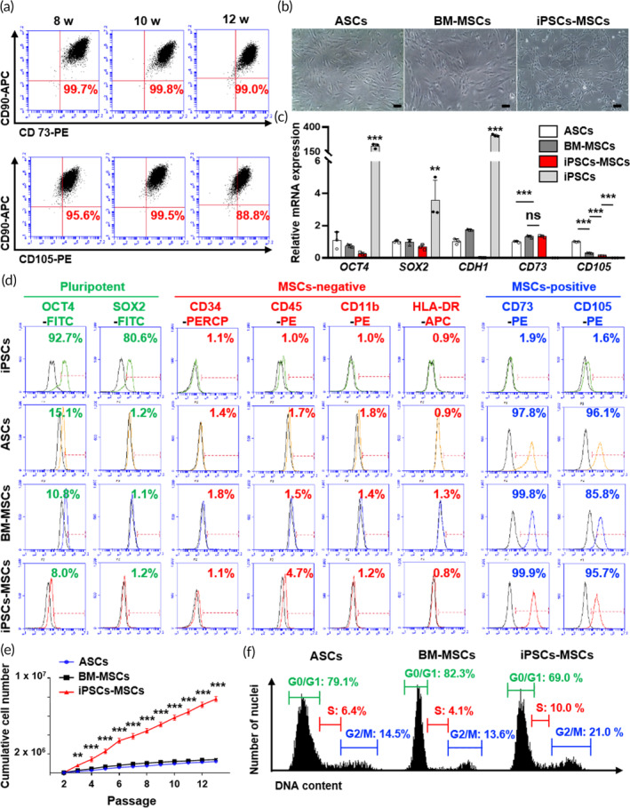
Characterization of condensed MSC mass‐derived mesenchymal stem cells (iPSCs‐MSCs). (a) Immunophenotype analysis of iPSCs‐MSCs isolated at 8, 10, and 12 weeks with MSCs markers CD73, CD90, and CD105 through flow cytometry. In every group, CD73 and CD90 double‐positive cells were higher than 99%. CD90 and CD105 double‐positive cells were approximately 90%. (b) Assessment of the morphology of ASCs, BM‐MSCs, and iPSCs‐MSCs via microscopy. Scale bar, 100 μm. (c) Comparison of mRNA expression of pluripotent markers (OCT4, SOX2, and CDH1) and MSC markers (CD73, CD105) between ASCs, BM‐MSCs, iPSCs‐MSCs, and iPSCs. The data are presented as the means ± s.d. (n = 3, biologically independent samples). *p < 0.05, **p < 0.01, ***p < 0.001, ns: no significance; based on one‐way ANOVA followed by Tukey's post hoc test. (d) Immunophenotype analysis of pluripotent markers (OCT4, SOX2), MSC‐negative markers (CD34, CD45, CD11b, and HLA‐DR), and MSC‐positive markers (CD73 and CD105) between iPSCs, ASCs, BM‐MSCs, and iPSCs‐MSCs through flow cytometry. (e) Measurement and comparison of the cumulative cell number until passage 13. The data are presented as the means ± s.d. (n = 3, biologically independent samples). *p < 0.05, **p < 0.01, ***p < 0.001, ns: no significance; based on one‐way ANOVA followed by Tukey's post hoc test. (f) Flow cytometry plot showing the cell cycle analysis of ASCs, BM‐MSCs, and iPSCs‐MSCs with DNA contents.
