
Figure 5.
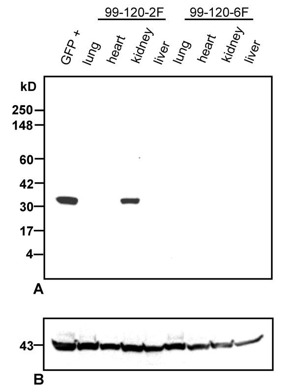
Western blot analysis of protein extracts from tissues of GUE-GFP transgenic mice. Tissue extracts from a transgenic mouse (99-120-2F) and a negative control mouse (99-120-6F) were separated on 4–20 % SDS-PAGE and transferred onto the membrane. The GFP positive control was from a cell line with GFP expression. 12 μg of total protein was loaded in each lane. A. Immunoreacting bands were detected using a polyclonal anti-GFP antibody (Clontech). B. The same blot was reprobed with a mouse monoclonal anti-actin antibody (Chemicon) to show that equal amount of protein was loaded in each lane.