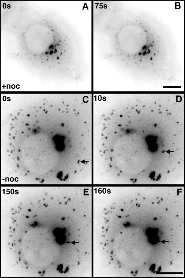
Figure 3.

Microtubule-dependent movement of peripheral EB1-ΔN2-GFP aggregates. Panels A and B. Single frames from additional file 4 showing a living cell expressing EB1-ΔN2-GFP after 2 h incubation in nocodazole. All EB1-ΔN2-GFP aggregates are immobile. Bar = 10 μm. Panels C-F. Single frames from additional file 5 showing a living cell expressing EB1-ΔN2-GFP during the recovery phase following nocodazole wash out. The retrograde transport of a peripheral aggregate and its incorporation into a large perinuclear aggregate is arrowed. Times shown are relative to the first frame in the sequence. Bar = 10 μm.
