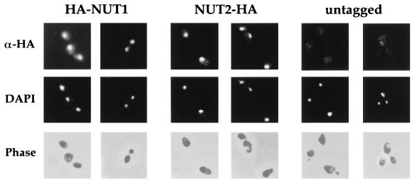
FIG. 5.
Nut1p and Nut2p are localized to nuclei. Cells bearing integrated copies of the HA-NUT1 fusion (pRKT542; columns 1 and 2), the NUT2-HA fusion (pRKT535; columns 3 and 4), or no fusion (columns 5 and 6) were harvested in mid-log phase, fixed, and stained with antibodies against the HA epitope (α-HA) as described in Materials and Methods. Localization of the tagged epitope was revealed by rhodamine-conjugated anti-mouse antibodies in the first row. For the same field of cells, DNA was visualized by 4′,6-diamidino-2-phenylindole (DAPI) staining in the second row. Cell outlines were visualized by phase-contrast microscopy in the third row.

