Figure 1.
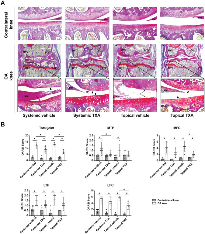
Systemic and topical TXA treatment attenuate articular cartilage degradation after ACLT. (A) Bone-Inflammation-Cartilage staining sections in the medial compartments of OA and contralateral knees at 4 weeks after ACLT (scale bar = 100 μm). Arrowheads indicate areas with loss of Safranin O staining. Arrows indicate clefts or erosion in the articular cartilage and irregular surface. (B) The OARSI scoring of the total joint, MTP, MFC, LTP, and LFC. The data are expressed as means ± SD; n = 5-7 per group as indicated (2-way analysis of variance followed by the Tukey post hoc test). *P < .05 for comparisons denoted by bar. ACLT, anterior cruciate ligament transection; LFC, lateral femoral condyle; LTP, lateral tibial plateau; MFC, medial femoral condyle; MM, medial meniscus; MTP, medial tibial plateau; OA, osteoarthritis; OARSI, Osteoarthritis Research Society International; TXA, tranexamic acid.

