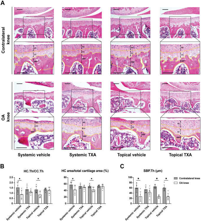
Figure 2.
Systemic and topical TXA treatment preserve hyaline cartilage after ACLT. (A) The representative histological images of the medial tibial cartilage stained with hematoxylin and eosin 4 weeks after ACLT (scale bar = 100 μm). A box (400 × 300 μm) was centered on the medial tibial plateau. The TM, CL, and LM of the SBP are marked by yellow dotted lines. The HC, CC, and SBP are labeled with black double-headed arrows. (B) Quantitative analysis of the ratio of HC thickness to CC thickness (HC.Th/CC.Th) and percentage of the HC area out of the total cartilage area. (C) Quantitative analysis of the SBP thickness (SBP.Th). All data are expressed as mean ± SD; n = 5-7 per group as indicated (2-way analysis of variance followed by the Tukey post hoc test). *P < .05 for comparisons denoted by bar. ACLT, anterior cruciate ligament transection; CC, calcified cartilage; CL, cement line; HC, hyaline cartilage; LM, lower margin; OA, osteoarthritis; SBP, subchondral bone plate; TM, tidemark; TXA, tranexamic acid.

