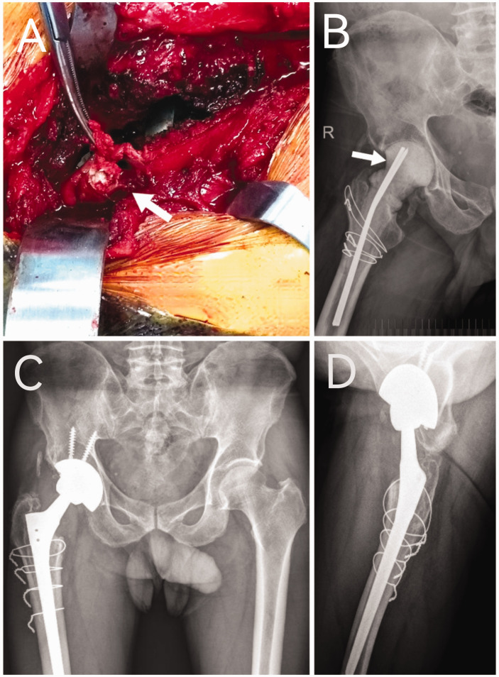
Figure 3.
Caseous tissue (white arrow) was seen in the supraacetabular area during the first-stage revision surgery (a) in a 53-year-old male patient that had undergone right total hip arthroplasty for alcoholic femoral head necrosis 2 years previously. A plain radiograph showing the bone cement spacer (white arrow) that was placed during the first-stage revision (b). Two-year postoperative follow-up plain pelvic anteroposterior (c) and lateral (d) radiographs of the right hip confirmed no manifest recurrence of infection. The colour version of this figure is available at: http://imr.sagepub.com.

