
Figure 2.
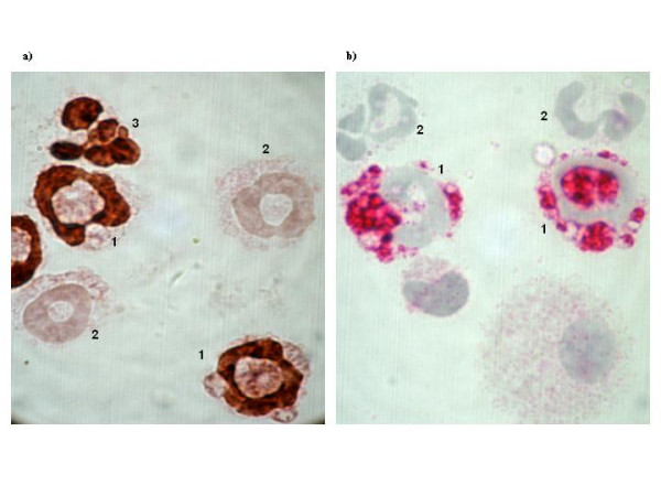
Photomicrographs (original magnification: ×1000) of BAL cells immunocytochemical analysis of staining positively for BrdU (brown nuclear, a) and CD34 (red, b) in sensitized and exposed to OVA on five consecutive days mice. a) 1- BrdU+ eosinophil, 2- BrdU- eosinophil, 3- BrdU+ neutrophil. b) 1- CD34+ granulocyte, 2- CD34- granulocyte.