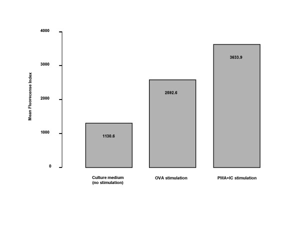
Figure 9.
Intracellular expression of IL-9, detected by flow cytometry in the bone marrow CD4+ cells from OVA-sensitized and exposed for 6 days animals, cultured in vitro with OVA or PMA (phorbol myristate acetate) + IC (calcium ionophore). Data are expressed as time-fold increase of the mean fluorescence intensity (MFI) of the baseline expression (culture with plain medium).

