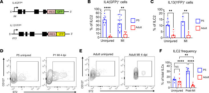
Figure 2. IL-4 and IL-13 expression is enriched in neonatal ILC2s.
(A) Schematic of IL-4GFPKI and IL-13YFPKI transgenic mouse line alleles. (B) Quantification of GFP+ cells in P5 versus adult mice in uninjured and at 4 dpi after MI. GFP+ cells in IL-4GFPKI/WT mice were normalized to WT age-matched littermates. (C) Quantifications of YFP+ cells in P5 versus adult mice in uninjured and at 4 dpi. YFP+ cells in IL-13YFPKI/WT mice were normalized to WT age-matched littermates. (D and E) Representative contour plots of CD127 versus IL-1RL1 in cardiac non–B/T lymphoid cells in neonatal (D) and adult (E) mouse hearts. (F) Quantification of ILC2 frequency in P5 versus adult mouse hearts either uninjured or at 4 dpi. Data are shown as mean ± SD. Each data point represents 1 mouse. **P < 0.01, ****P < 0.0001. Age effect by 2-way ANOVA and Sidak’s post hoc test for uninjured and after MI conditions in B and C. Interaction effect of age and condition by 2-way ANOVA and Sidak’s post hoc test in F.

