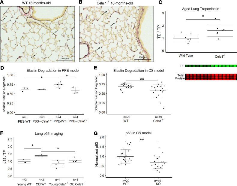
Figure 3. Lung elastin degradation and senescence in 3 mouse emphysema models.
(A) Photomicrographs of Hart-stained lung sections of aged WT and (B) Cela1–/– mouse lung show both preservation of alveolar structure in Cela1–/– mice and preservation of alveolar septal tip elastin bands (arrows). Scale bars: 100 μm. (C) By Western blot, the lungs of Cela1–/– mice have a greater amount of total lung tropoelastin (TE). Normalization is by total protein (TP) and comparison is by 2-tailed Welch’s t test. *P < 0.05. n = 7 per group. (D) At 42 days after PPE, the lungs of WT mice had more degraded elastin than PBS-treated WT mice and PPE-treated Cela1–/– mice. P < 0.05 by 1-way ANOVA and Holm-Šidák post hoc comparisons are shown by *P < 0.05. Number of specimens per group is noted by n. (E) After 8 months of exposure, the lungs of cigarette smoke–exposed (CS-exposed) Cela1–/– mice had less degraded elastin than the lungs of WT mice. (F) p53 levels were increased in aged WT but not Cela1–/– lungs. *P < 0.05 by 2-tailed Welch’s t test. (G) The lungs of CS-exposed Cela1–/– mice had less p53 than the lungs of WT mice. **P < 0.01 by 2-tailed Welch’s t test.

