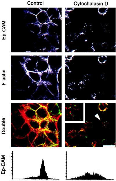
FIG. 1.
Effect of cytochalasin D on the actin cytoskeleton and the subcellular localization of Ep-CAMs in human epithelial RC-6 cells. Control cells and cells treated for 2 h with 10 μg of cytochalasin D per ml were stained for either Ep-CAM with MAb 323/A3 (green fluorescence) or polymerized actin filaments with phalloidin-TRITC (red fluorescence). Note the substantial disappearance of the Ep-CAMs from the cell-cell boundaries after the treatment. Internalized Ep-CAM does not colocalize with actin patches in the treated cells in the cytochalasin D-treated, double-stained cells (note the cell marked by an arrowhead and presented at larger magnification in the upper left corner). The internalization of Ep-CAM was verified by staining with MAb 323/A3 of living RC-6 cells (flow cytometry histograms below). Bar, 25 μm.

