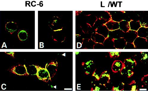
FIG. 8.
Colocalization of Ep-CAM and α-actinin in RC-6 (A, B, and C) and L (D and E) cells. Double-immunofluorescence staining was performed with fixed RC-6 and L cells with MAb 323/A3 for Ep-CAM (red fluorescence) and MAb CB-11 for α-actinin (green fluorescence). The L cells shown in panel E were cultured for 2 h in the presence of 10 μg of cytochalasin D per ml prior to fixation. Note the absence of colocalization between the two molecules in single cells and also in the areas of adhesion plaques (arrows), as well as in L cells treated with cytochalasin D. Bars, 10 μm (A, B, and C) and 5 μm (D and E).

