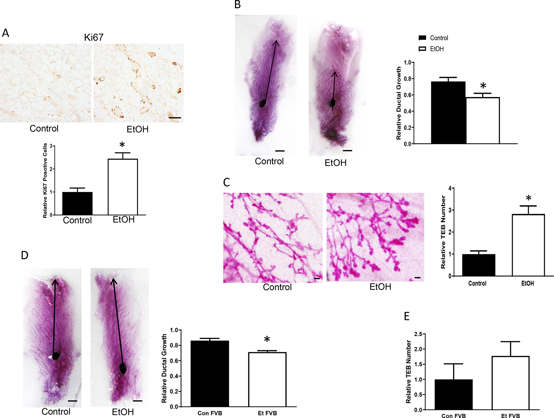
Fig. 5.

Alcohol’s effects on the development of mammary glands. Adolescent MMTV-Wnt1 mice received chronic alcohol exposure through liquid diet for 5 weeks as described above. A. Mammary glands were dissected and analyzed for Ki67 immunohistochemistry (IHC). Ki67-positive cells were counted and expressed relative to controls, n = 7 for each group. * significant difference from controls, p < 0.05. Bar = 25 μm. B. Mammary glands were collected for whole mount staining. Images were captured by a camera, and ductal growth was determined as the distance from the lymph node to the farthest point of the longest duct and expressed relative to the distance to the farthest limit of the mammary fat pad. n = 7 for each group. * significant difference from controls, p < 0.05. Bar = 5 mm. C: Terminal end buds (TEB) were counted and quantified relative to the controls, n = 7 for each group. * significant difference from controls, p < 0.05. Bar = 100 μm. D. Adolescent wild type (WT) FVB mice were exposed to alcohol through liquid diet for 5 weeks as described above. The ductal growth was determined as described above. n = 5 for each group. * significant difference from controls, p < 0.05. E. Adolescent WT FVB mice received chronic alcohol exposure through liquid diet for 5 weeks as described above, and relative TEB number was determined.
