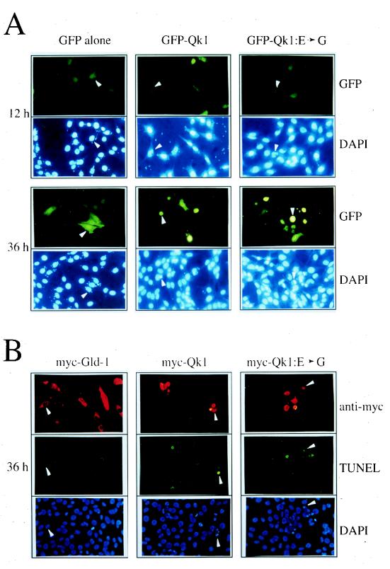
FIG. 6.
Qk1 and Qk1:E➛G induce apoptosis in NIH 3T3 cells. (A) NIH 3T3 cells were transfected with an expression vector encoding GFP alone, GFP-Qk1, or GFP-Qk1:E➛G. After 12, 24, 36, and 48 h, the cells were fixed and stained with DAPI to visualize the nuclei. The top photograph in each pair shows the fluoresceinated cells containing GFP, and the lower photograph shows the DAPI-stained nuclei. The white arrowheads were used to align the top and bottom photographs. (B) NIH 3T3 cells were transfected with expression vector encoding myc–GLD-1, myc-Qk1, or myc-Qk1:E➛G. The myc epitope-tagged proteins were visualized by indirect immunofluorescence with a rhodamine-conjugated secondary antibody (anti-myc). The apoptotic cells were visualized by TUNEL with fluorescein-containing nucleotides (TUNEL), and the nuclei were stained with DAPI (DAPI). The three photographs each for myc–GLD-1, myc-Qk1, and myc-Qk1:E➛G represent the same field of cells as visualized with different filters. The white arrowheads were used to orient the cells in the photographs.

