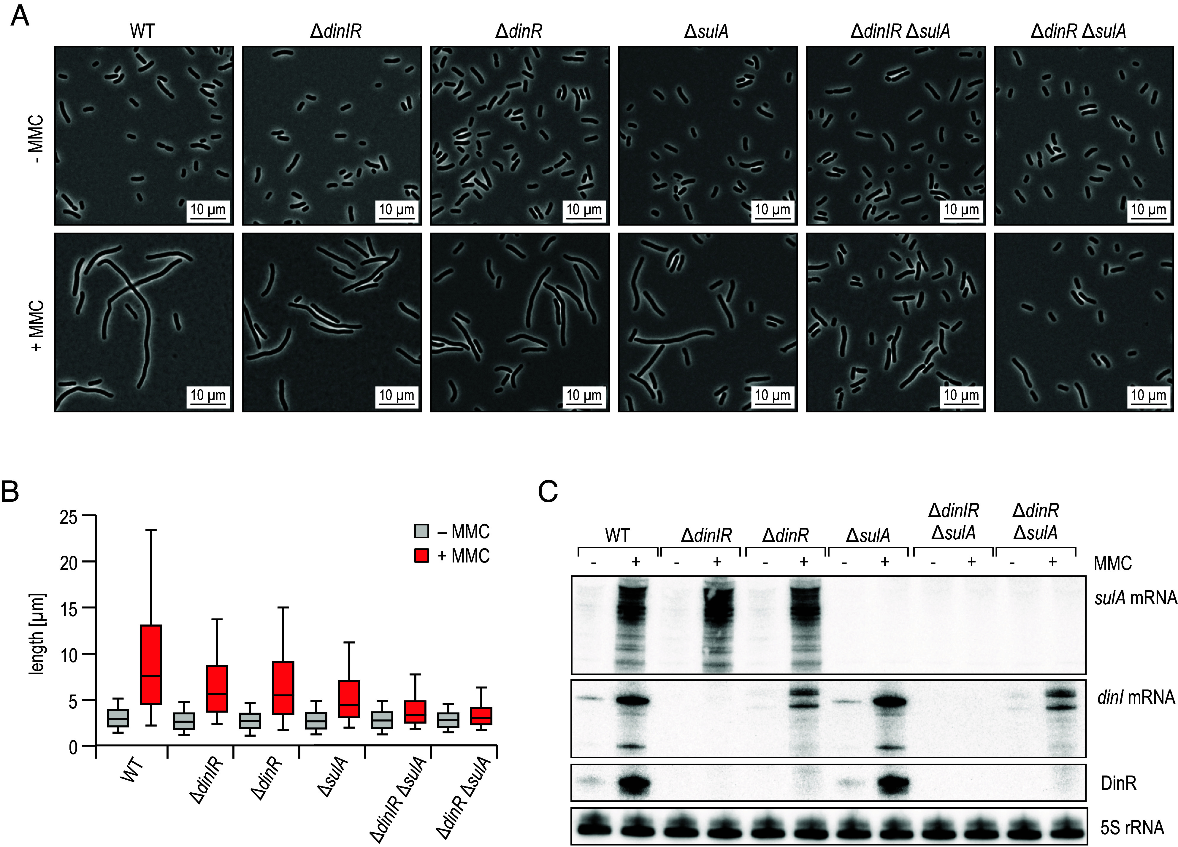
Fig. 6.

DinR and SulA contribute to cell filamentation in response to DNA damage. (A) K. pneumoniae WT, ΔdinIR, ΔdinR, ΔsulA, ΔdinIR ΔsulA, and ΔdinR ΔsulA cells were diluted from overnight cultures into fresh medium, and grown for 30 min. Cultivation was continued for 5 h in the presence (+) or absence (−) of MMC to induce DNA damage. Cell morphology was assessed by phase contrast microscopy. Details on mutant design at the dinIR locus are provided in SI Appendix, Fig. S12A. (B) Analysis of cell lengths in samples described in (A). The center line indicates the median, boxes represent the 25th and 75th percentiles, and lower and upper whiskers represent the 10th and 90th percentiles, respectively. (C) RNA samples were collected from cells cultivated as described in (A) for 30 min in the presence (+) or absence (−) of MMC.
