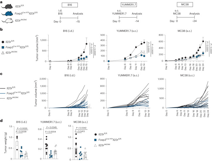
Fig. 2. Treg cells mediate the tumor-promoting functions of IL-23.
a–d, Il23rfl/fl, Foxp3Cre-YFPIl23rfl/fl and Il23rdel/del mice were inoculated i.d. with B16 tumor cells, inoculated subcutaneously (s.c.) with YUMMER1.7 tumor cells or inoculated s.c. with MC38 tumor cells, and tumors were analyzed around days 15, 14 and 24 after inoculation. The data show the results of three independent experiments (B16: n = 3 Il23rfl/fl mice, n = 5 Il23rdel/del mice, n = 8 Foxp3Cre-YFPIl23rfl/fl mice; MC38: n = 12 Il23rfl/fl mice, n = 7 Foxp3Cre-YFPIl23rfl/fl mice, n = 7 Il23rdel/del mice; YUMMER1.7: n = 10 Il23rfl/fl mice, n = 7 Foxp3Cre-YFPIl23rfl/fl mice, n = 8 Il23rdel/del mice). a, Schematic illustration of the experimental approach. b, Tumor volume kinetics of the experimental groups measured by caliper gauge. Data are shown as mean ± s.e.m. Statistical significance was determined by two-way analysis of variance (ANOVA) with a Sidak’s post hoc test. c, Tumor volume kinetics of individual mice measured by caliper gauge. d, Bar graph displaying the final tumor weight. Data are displayed as mean ± s.e.m. Statistical significance was determined using two-tailed t-tests.

