FIG. 4.
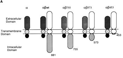
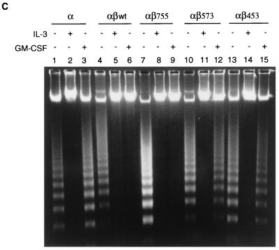
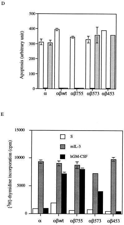
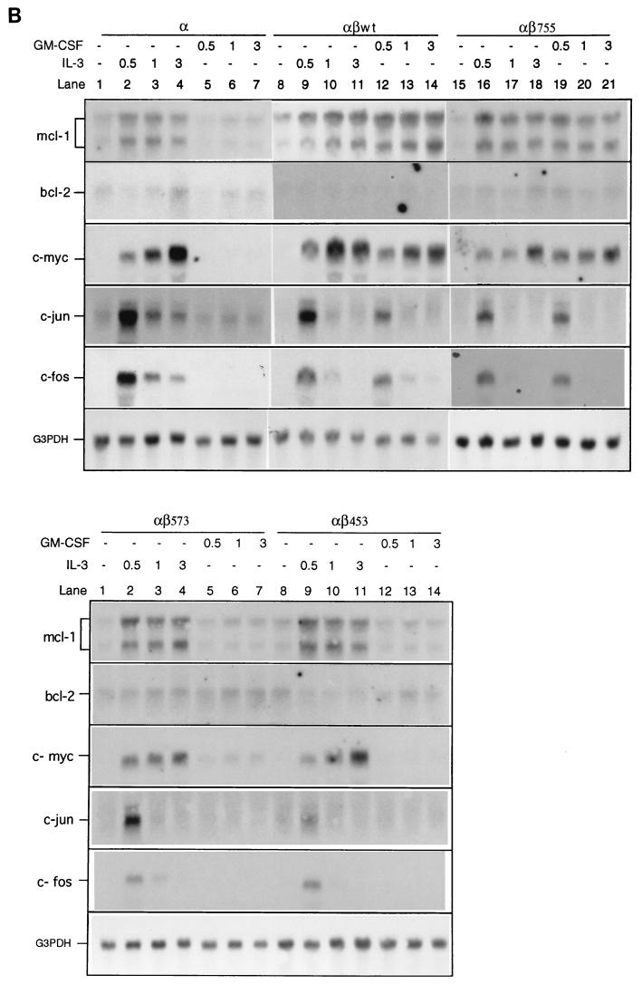
Mcl-1 induction requires the membrane-distal domain of the GM-CSF receptor β chain. (A) Schematic representation of human GM-CSF receptor mutants transfected into Ba/F3 cells. (B) Ba/F3 cells expressing various receptor mutants were depleted of cytokine for 20 h before they were stimulated with mIL-3 or hGM-CSF. At various times after stimulation (0.5 to 3 h), the cells were lysed and their total RNA was analyzed by Northern blotting with specific probes as indicated in the figure. (C) Equal numbers (2 × 10⁶) of Ba/F3 cells expressing various GM-CSF receptor mutants were seeded for 24 h in 10 ml of medium containing no cytokine, mIL-3, or hGM-CSF, and then their genomic DNA was extracted and analyzed by agarose gel electrophoresis (2% agarose). (D) Cells treated as in panel C were stained with biotinylated annexin-V and Texas red-conjugated streptavidin as described in Materials and Methods. The positively stained (apoptotic) cells were quantified with Cytofluor 2350. The fluorescence units of the annexin-V-bound cells are plotted here to reflect the absolute numbers of apoptotic cells. □, S; , mIL-3; , hGM-CSF. (E) Mitogenic activity of Ba/F3 cells expressing various receptor mutants. Cells (104) cultured in medium containing no cytokine (S), mIL-3 or hGM-CSF were pulse-labeled with [3H]thymidine for 20 min and lysed, and the incorporated counts were measured with a β-counter.


