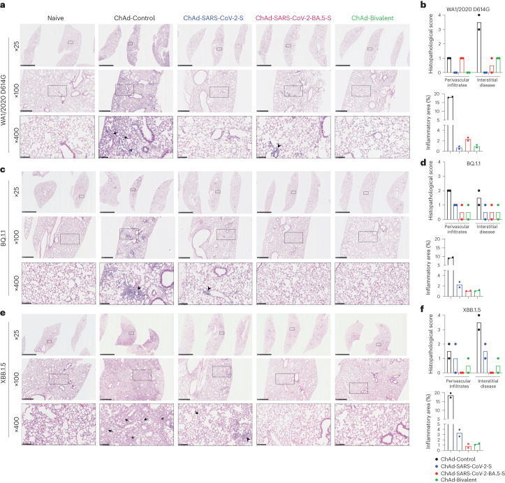
Fig. 5. ChAd vaccines protect mice against SARS-CoV-2-induced lung pathology.
Seven- to 9-week-old female K18-hACE2 mice were immunized with ChAd vaccines and challenged with WA1/2020 D614G, BQ.1.1 or XBB.1.5, as described in Fig. 1b. Hematoxylin and eosin staining of lung sections (a, c and e) and corresponding pathology scores (b, d and f) using two different analysis modes (see Methods) are shown. Data for WA1/2020 D614G (a and b), BQ.1.1 (c and d) and XBB.1.5 (e and f) are shown. Images show low-power (×25; scale bars, 2.5 mm), medium-power (×100; scale bars, 500 µm) and high-power (×400; scale bars, 100 µm) magnifications. Images at high magnifications (×400) show infected lungs with immune cell infiltrates (arrows), thickened septa and consolidated air space (asterisks) and foci of immune cells near perivasular and peribronchial spaces (arrow heads). Images are representative of n = 2 mice per group.

