
Fig. 5.
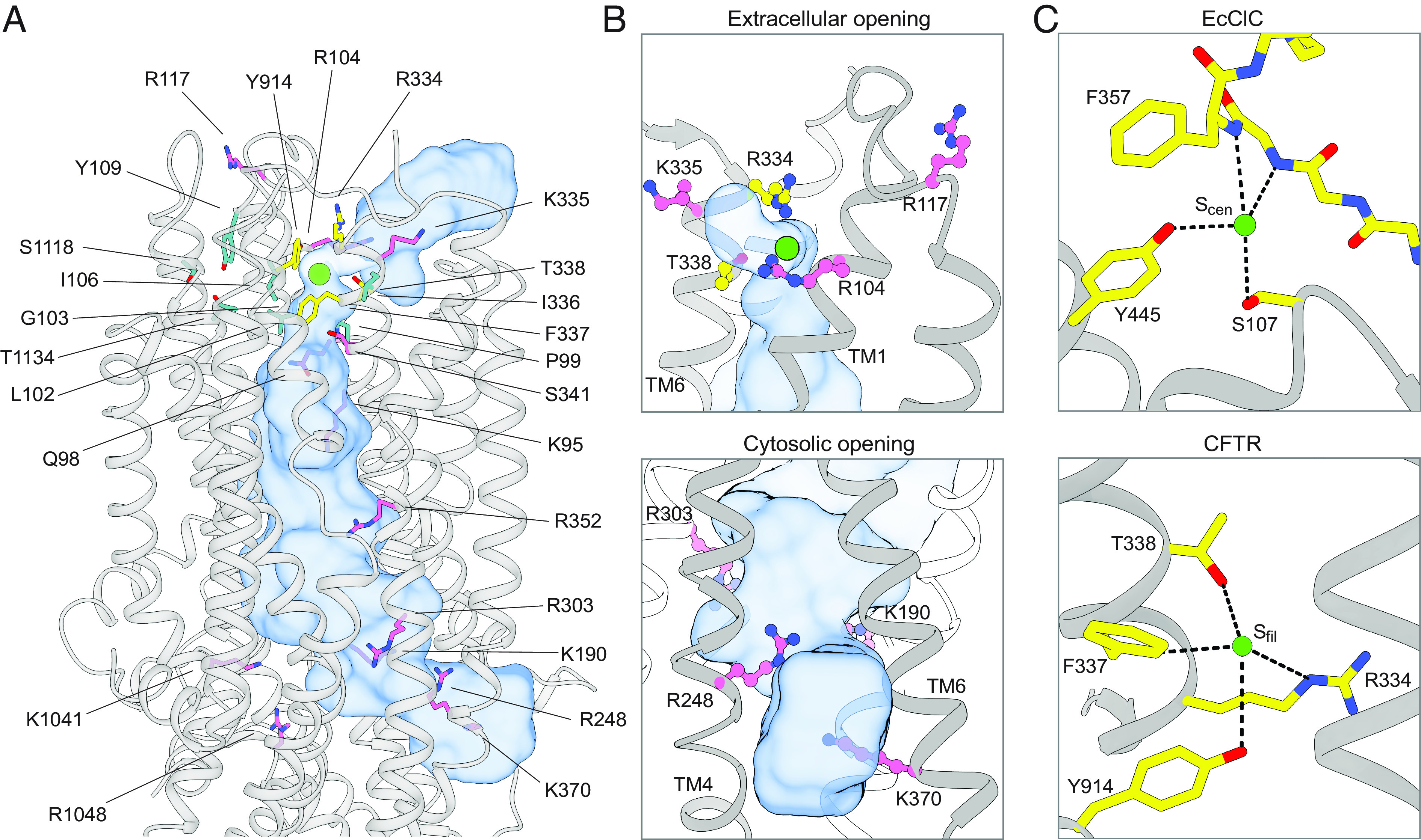
CFTR residues that contribute to ion selectivity and/or ion conduction. (A) Residues shown to contribute to ion selectivity or conduction in the literature are mapped onto the three-dimensional structure of CFTR. The pore is shown as a blue surface. Chloride is shown as a green sphere. Residues in the inner and outer vestibules are pink. Residues that directly coordinate chloride at Sfilter are yellow. Residues of the second coordination sphere of Sfilter are cyan. (B) Closeup views of the extracellular opening (Upper) and cytosolic opening (Lower) of CFTR’s pore. The pore is outlined as a blue surface. (C) Chloride coordination at the Scen chloride-binding site in wild-type EcClC (Upper, Protein Data Bank 1OTS) (80) and at the Sfilter chloride-binding site in CFTR (Lower).