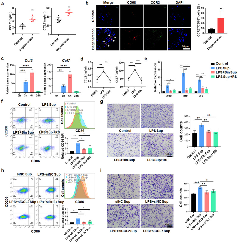
Fig. 2. Degenerative NPCs induce the recruitment and proinflammatory polarization of MΦs through the CCL2/7-CCR2 axis.
a ELISA analysis of the levels of CCL2 and CCL7 in human NP tissue. b Immunofluorescence results showing the distribution of CCR2+ and CD68+ cells in human NP tissues. c qPCR results showing Ccl2 and Ccl7 gene expression in rNPCs treated with LPS (1 µg/ml) for 0/3/6/24 h. d ELISA results showing the levels of CCL2 and CCL7 in the supernatant of rNPCs treated with bindarit for 1 h and LPS for 6 h. e qPCR results showing inflammation-related genes in rMΦs induced by supernatant from rNPCs treated with bindarit for 1 h and LPS for 6 h. f FCM analysis and quantification of CD68+CD11b+CD86+ and CD68+CD11b+CD206+ rMΦs as determined by geomean fluorescence intensity (GFI). The supernatant was extracted from rNPCs that were pretreated with bindarit for 1 h and LPS for 6 h with or without RS102895 for 1 h. g Transwell assays were used to detect the migration of rMΦs in the different groups. h rMΦs were treated with the supernatant extracted from LPS-treated rNPCs with CCL2 and CCL7 knockdown. The quantification of CD68+CD11b+CD86+ and CD68+CD11b+CD206+ rMΦs was detected by FCM. i Transwell assays were used to detect the cell migration of rMΦs in the different groups. Experiments were performed at least 3 times, and the data are presented as the means ± SDs. *p < 0.05; **p < 0.01; ***p < 0.001; ns, not significant, ANOVA. Con, control; Sup, supernatant; L, LPS; Bin, bindarit; RS, RS102895.

