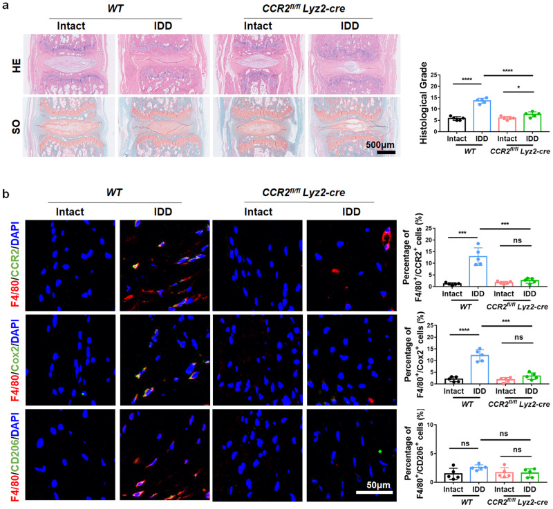
Fig. 8. CCR2 knockout in MΦs inhibits MΦ infiltration and IDD.
a HE and SO staining of CCR2fl/flLyz2-cre and WT mouse IVD tissue. b Immunofluorescence images and quantitative analysis of F4/80+/CCR2+, F4/80+/Cox2+ and F4/80+/CD206+ double-positive cells in the IVD tissue of CCR2fl/flLyz2-cre and WT mice. Experiments were performed 5 times independently, and the data are presented as the means ± SDs. *p < 0.05; **p < 0.01; ***p < 0.001; ns: not significant, ANOVA.

