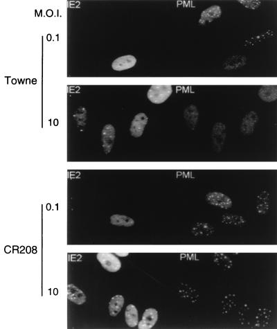
FIG. 1.
Cells infected with an HCMV mutant that does not express IE1 fail to disperse the PML protein from punctate domains (PODs) into a nuclear diffuse pattern. The photographs show a comparison of PML staining patterns in HF cells infected with either HCMV(Towne) (upper two panels) or the CR208(ΔIE1) virus (lower two panels) at a low MOI of 0.1 and a high MOI of 10. At 6 h after infection, the cells were fixed with paraformaldehyde followed by double-label IFA. IE2 was detected with mouse MAb 12E2 and FITC-labeled anti-mouse IgG (left). PML was detected in the same fields with rabbit anti-PML PML-C PAb and rhodamine-coupled anti-rabbit IgG (right). Note the typical punctate POD patterns of PML in adjacent uninfected cells at low MOI.

