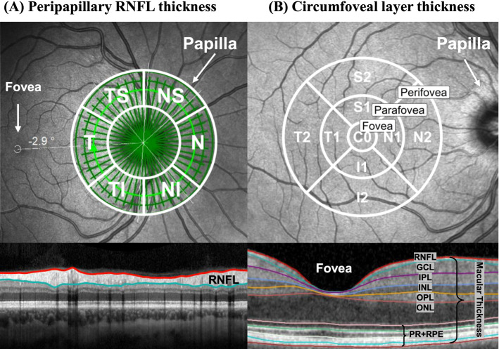
Fig. 1.
Schematic overview of OCT examinations. A Peripapillary RNFL thickness from the 3 circle scans with the 6 sector Garway-Heath grid (white circles and lines) (Garway-Heath et al., 2000). B Circumfoveal macular and intraretinal layer thickness measured within the 9 sector ETDRS grid (white circles and lines) (Chew, 1996). Sector data were summarized into total thickness and in 3 foveal regions: fovea, parafovea and perifovea. Abbreviations: GCL = ganglion cell layer; I = inferior; INL = inner nuclear layer; IPL = inner plexiform layer; N = nasal; NI = nasal-inferior; NS = nasal-superior; ONL = outer nuclear layer; OPL = outer plexiform layer; RNFL = retinal nerve fiber layer; PR + RPE = photoreceptor-retinal pigment epithelium complex; S = superior; T = temporal; NI = temporal-inferior; TS = temporal-superior (Friedel et al., 2022a)

