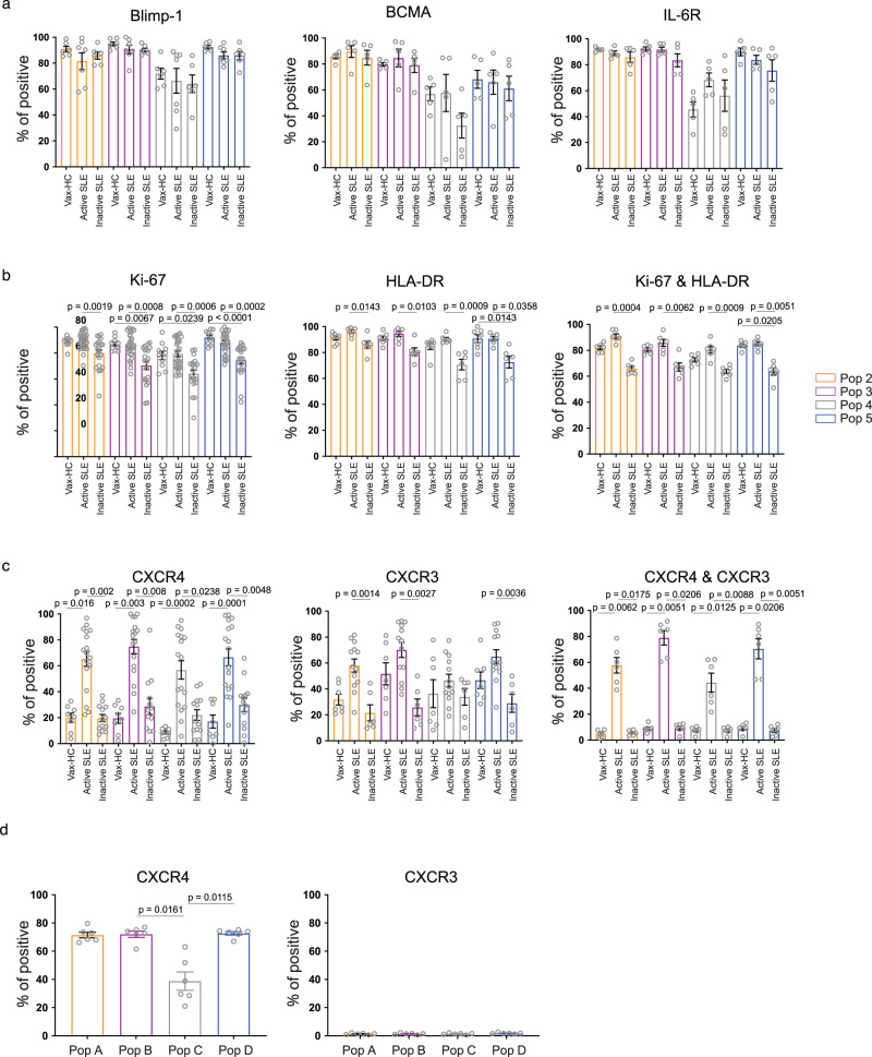
Fig. 3. Immune phenotype of circulating ASC in active SLE.
a–c Peripheral blood mononuclear cells (PBMCs) were isolated from steady-state healthy donors, influenza vaccinated heathy subjects on day 7 post immunization, active SLE patients, or inactive SLE patients and isolated cells were stained and analyzed by flow cytometry. a Expression of intracellular molecular Blimp-1 in ASC populations from vaccinated heathy subjects (n = 6), active SLE patients (n = 7), or inactive SLE patients (n = 6); and surface molecules BCMA (n = 5 per group) and IL-6R (n = 5 per group). Data are shown as mean ± SEM. b Expression of intracellular Ki-67 in ASC populations from vaccinated heathy subjects (n = 10), active SLE patients (n = 28), or inactive SLE patients (n = 21), surface HLA-DR on ASC populations from vaccinated heathy subjects (n = 7), active SLE patients (n = 6), or inactive SLE patients (n = 6) and their co-expression on ASC populations (n = 6). Data are shown as mean ± SEM. c Expression of surface expression of CXCR3 from vaccinated heathy subjects (n = 7), active SLE patients (n = 13), or inactive SLE patients (n = 6); CXCR4 from vaccinated heathy subjects (n = 8), active SLE patients (n = 17), or inactive SLE patients (n = 13), and their co-expression on ASC populations (n = 6). Data are shown as mean ± SEM. d Bone Marrow mononuclear cells (BMMCs), were isolated from SLE patients, and analyzed by flow cytometry for the expression of CXCR4 (left) and CXCR3 (right) on BM PC populations, Pop A-D, from SLE patients (n = 6). Data are shown as mean ± SEM. Statistical significance was assessed using Kruskal-Wallis test followed by Dunn’s test for multiple comparisons within the same ASC population (a-d). p values are shown on plots. Source data are provided as a Source Data file.

