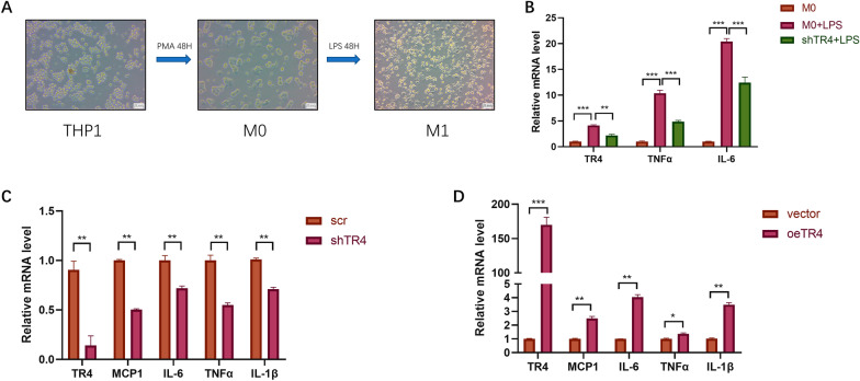
Fig. 5.
TR4 promotes inflammatory factor secretion during urosepsis. A THP-1 cells can be induced to differentiate into M0 macrophages after treatment with 100 ng/ml PMA for 48 h, and M0 macrophages, which are round or oval in shape, can be observed under a microscope. The M0 cells were then treated with 500 ng/ml LPS for 48 h to induce M1 macrophages. Under the microscope, irregular cell morphology and obvious cell protrusions could be observed. B After LPS stimulation, the expression levels of TR4, TNFα, and IL-6 in macrophages were significantly increased. When TR4 was knocked down, the expression levels of TR4, TNFα, and IL-6 in macrophages decreased. *p < 0.05, **p < 0.01, ***p < 0.001. C After knocking down TR4, the expression levels of MCP1, IL-6, TNFα, and IL-1β were significantly decreased; **p < 0.01. D After overexpressing TR4, the expression levels of MCP1, IL-6, TNFα, and IL-1β were significantly increased; *p < 0.05, **p < 0.01, ***p < 0.001

