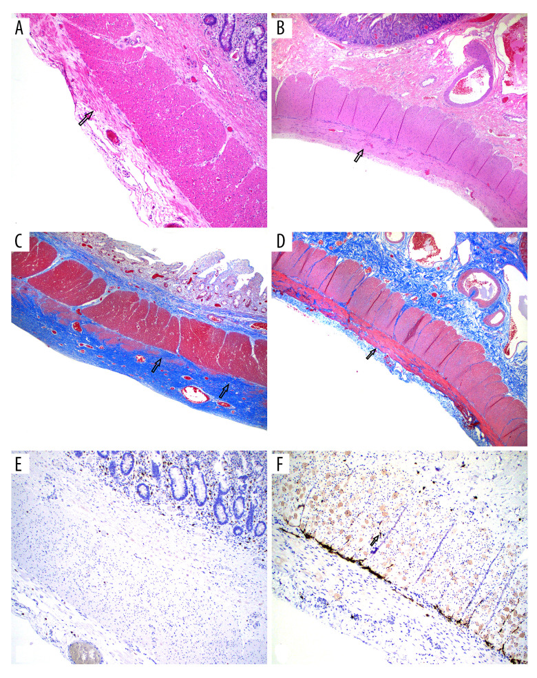
Figure 1. Native intestine.
(A) H&E-stained full-thickness native jejunum showing thinned external layer of muscularis propria with muscle loss and fibrosis on the left (arrow points to remaining bundle of muscle in the midst of fibrosis). (B) Normal intestine biopsy for comparison showing normal external layer of muscularis propria (100×). (C) Trichrome stain showing replacement of most of the external layer of muscularis propria by fibrosis (blue color, arrows). (D) Normal muscularis propria for comparison. (40×). (E) CD117 stain for interstitial cells of Cajal (stain cells brown to black, arrow) showing marked reduced number of cells in the myenteric plexus and inner circular layer (100×). (F) normal distribution of cells of Cajal for comparison.

