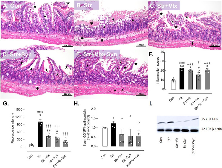
Figure 3. Histomorphological, intestinal barrier-related function and neuroprotective protein profiles of ileum as determine by H&E stating, fluorescence assay and Western blotting analysis.
Histological images of ileum (A–E) and inflammatory scores in male rats under stress exposures (F). The morphology of the ileum revealed desquamation of the villi (black star), villous edema (black triangle), loss of villi (asterisks) and layer separation (black arrow). 70-kDa FITC-dextran intestinal permeability (G), relative glial cell-derived neurotrophic factor (GDNF) protein in ileum in stressed rats (H) and the representative expression of GDNF beta actin (I). *p < 0.05, **p < 0.01 and ***p < 0.001 compared to vehicle-treated control rats. †p < 0.05 and †††p < 0.001 compared to vehicle-treated stressed rats (n = 4 rats/group). Con, control; Str, stressed+vehicle; Str+Vlx, stressed+venlafaxine; Str+Syn, stressed+synbiotic supplement; Str+Vlx+Syn, stressed+venlafaxine+synbiotic supplement.

