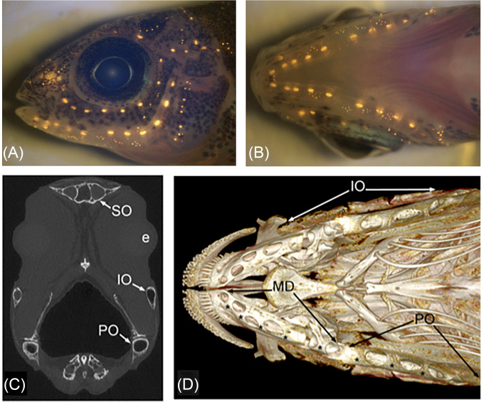
FIG. 6.
(Color online) Neuromasts in the peacock cichlid, Aulonocara stuartgranti in (A) lateral and (B) ventral views as revealed by vital fluorescent staining of hair cells in neuromasts (with 4-Di-2-ASP). CNs are larger than SNs. (C) Transverse μCT (micro-computed tomographic) slice through the head of adult Aulonocara baenschi at the level of the lens of the eye (e), indicating the lumen of the preopercular (PO), infraorbital (IO), and supraorbital (SO) canals. (D) 3-D reconstructions (μCT) of cranial skeleton, in ventral view, showing the mandibular (MD), preopercular (PO), and infraorbital (IO) LL canals in adult Aulonocara baenschi. Asterisks (*) indicate the location of CNs within the MD canal, found within the dentary and anguloarticular bones of the mandible, and in the PO canal in the ventral portion of the L-shaped preopercular bone. (A) and (B) Reprinted from Figs. 2(E) and 2(F) from Becker, Bird, and Webb, “Post-embryonic development of canal and superficial neuromasts and the generation of two cranial lateral line phenotypes,” J. Morphol. 277(10), 1273–1291 (2016). Copyright 2016 Wiley Periodicals, Inc., John Wiley and Sons. (C) and (D) Reprinted from Figs. 3(D) and 3(C) from Webb, Bird, Carter, and Dickson, “Comparative development and evolution of two lateral line phenotypes in Lake Malawi cichlids,” J. Morphol. 275(6), 678–692 (2014). Copyright 2014 Wiley Periodicals, Inc., John Wiley and Sons.

