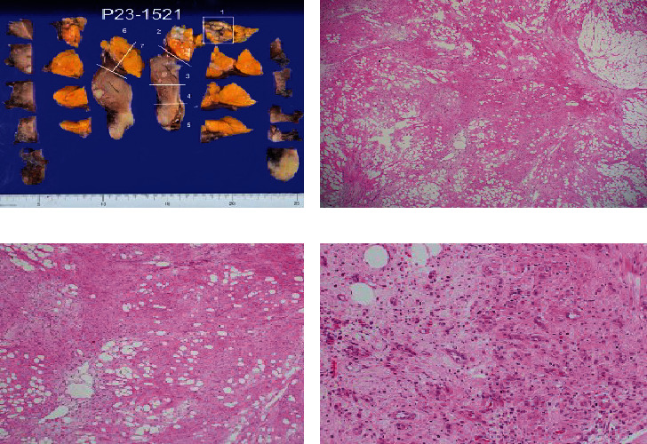
Figure 4.

Macroscopic findings and hematoxylin–eosin (HE) staining findings: (a) display of the pathological specimen site after ALTFiX fixation; (b) HE staining of specimen preparation site 1 (×100); (c) HE staining of specimen preparation site 1 (×200); (d) HE staining of specimen preparation site 1 (×400). Differences in the sizes of adipocytes and stromal cells were noted (b, c). There were irregularities in the nuclear findings of stromal cells and irregularities and thickening of the nuclear margins. Nuclear enrichment was observed (d).
