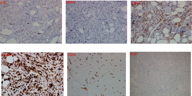
Figure 5.

Immunohistochemical staining: (a) p16 (×400). Some cytoplasm and nuclei were stained; (b) MDM2 (×400). A small part of the cytoplasm and nucleus was stained; (c) CDK4 (×400). The cytoplasm of long spindle-shaped cells of the connective tissue and the cytoplasm of adipocytes were stained; (d) desmin (×400). The cytoplasm of long spindle-shaped cells of the connective tissue and the cytoplasm of adipocytes were stained; (e) CD34 (×400). Some cell membranes were stained; (f) Ki-67 index = 1%.
