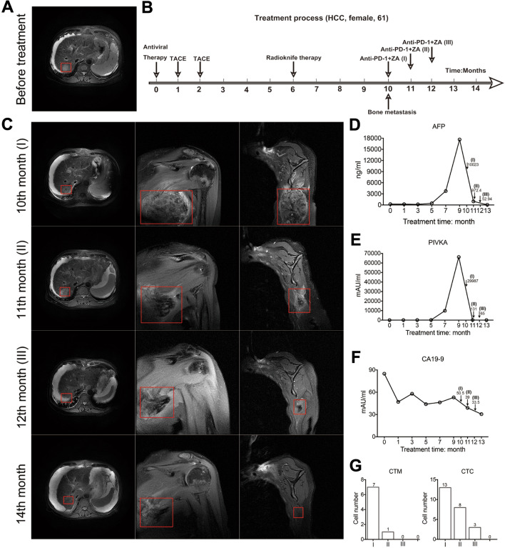
Fig. 1.
The case of combined ZA and anti-PD-1 in the treatment of bone metastasis from liver cancer. A MRI of the primary lesion of the liver cancer before treatment. B The process of treatment. (Antiviral therapy was given within the first month after the diagnosis of primary liver cancer.) The patient received two TACE treatments in the first and second month. At the 6th month, radioknife treatment was used for the patient. Bone metastasis was found at the 10th month. The anti-PD-1 treatments combined with ZA were applied in the 10th, 11th and 12th months, respectively. C MRI images of the liver and left scapula after the patient was found to have bone metastasis. At the 11th month, the left scapular lesion was significantly reduced on the 7th day after treatment. At the 12th month, the lesion in the right lobe of the liver was stable, and the metastasis in the left scapula was significantly reduced. At the 14th month, there was no progression of the intrahepatic lesion, and the extrahepatic metastasis almost disappeared. D, E High AFP and PIVKA were detected and subsequently reaching maximum values when bone metastasis was observed. Both of them decreased gradually after anti-PD-1 combined ZA treatment. F The patient had a high level of CA19-9 at diagnosis. From the beginning to the tenth month, the level of CA19-9 gradually decreased but remained above normal. And the rate of decrease gradually slowed down or even slightly increased. After the third administration of anti-PD-1 + ZA, CA19-9 decreased to the normal range (< 37mAU/ml). G With the application of the combined treatment, CTC and CTM were gradually reduced to 0

