Fig. 7.
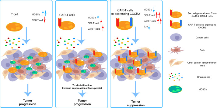
Graphic abstract. The left figure: In patients with pancreatic cancer, T cells demonstrate a capacity for infiltrating the pancreatic cancer microenvironment. However, this ability is hindered by the fibrotic nature of pancreatic cancer. Concurrently, MDSCs inhibit these infiltrating T cells, diminishing their tumor-destroying capabilities and ultimately facilitating tumor progression. The central figure: After administering CAR-T cells to patients with pancreatic cancer, these CAR-T cells possess a superior ability to infiltrate and recognize tumor cells compared to standard T cells. Nonetheless, the immunosuppressive network established by MDSCs alongside other suppressive cells within the immune microenvironment of pancreatic cancer remains unaltered. As a result, CAR-T cells are prevented from executing their normal tumor-killing function. The right figure: The introduction of CXCR2 into CAR-T cells led to the creation of CXCR2 CAR-Ts that inhibited the migration of MDSCs and CXCR2 macrophages to the pancreatic cancer site. This resulted in lower levels of IL-8 in the microenvironment, enabling CAR-T cells to enjoy enhanced infiltration abilities. This process also improved the composition of cells and factors within the pancreatic cancer microenvironment, thereby alleviating, to a certain degree, the immunosuppressive environment of pancreatic cancer. This ultimately resulted in the inhibition of in situ tumor formation and metastasis

