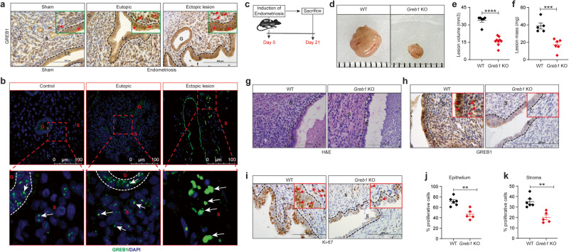
Fig. 7. GREB1 is required for endometriotic lesion growth in mice.
a-b, Representative images of GREB1 localization in mouse eutopic endometrium and ectopic lesion, (n = 5) (a) and human eutopic endometrium and ectopic lesion (n = 10 control, n = 10 eutopic and n = 10 ectopic lesion) (b). Red/White arrows, GREB1-positive cells. c Experimental timeline and procedure. Ectopic endometriotic lesion representative images (d), volumes WT (n = 6) and Greb1 KO (n = 9) (e), and masses WT (n = 5) and Greb1 KO (n = 6), f from mice 21 days after surgical induction of endometriosis. Paired, two-tailed, t-test. Data are presented as the mean ± SEM. *P < 0.05, **P < 0.01, ***P < 0.001 and ns non-significant. Representative images of ectopic lesions from WT and Greb1 KO mice stained with Hematoxylin and Eosin (g), anti-GREB1 antibody (h), and anti-Ki-67 antibody (i); red arrows, indicates respective positive cells (n = 5). Graphs display percentage of Ki-67-positive cells in endometriotic lesion epithelium (j), and stroma (k) from WT (n = 6) and Greb1 KO mice (n = 5). E epithelium, G gland, LE luminal epithelium, S stroma. Paired, two-tailed, t-test. Data are presented as the mean ± SEM. *P < 0.05, **P < 0.01, ***P < 0.001 and ns non-significant.

