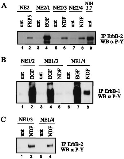
FIG. 2.
Ligand-induced tyrosine phosphorylation of ErbB-1 and ErbB-2. The different NIH sublines were starved for 18 h in serum-free medium and were left untreated (unt) or stimulated with the indicated growth factors (1 nM) or MAb FRP5 (10 μg/ml) for 10 min at room temperature. ErbB-1 was immunoprecipitated (IP) with a mixture of ErbB-1-specific MAbs EGFR1 and 528 (B); ErbB-2 was immunoprecipitated with 21N antiserum (A and C). The immune complexes were subjected to SDS-PAGE (7.5% gel) and analyzed by Western blotting (WB) using a phosphotyrosine-specific MAb (α P-Y). (C) Long exposure which enabled detection of tyrosine-phosphorylated endogenous ErbB-2.

