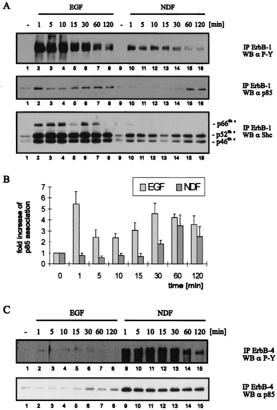
FIG. 8.
Time course of ligand-induced ErbB receptor activation and association with Shc and p85. NE1/4 cells were serum starved for 18 h and, prior to lysis, stimulated with 1 nM EGF or NDF (A and C) for the indicated periods at 37°C. Equal amounts of protein were immunoprecipitated (IP) with ErbB-1-specific MAbs EGFR1 and 528 (A) and ErbB4-specific polyclonal antibody C18 (C), and immune complexes were resolved by SDS-PAGE (8% gel). The membranes were probed by Western blotting (WB) with a phosphotyrosine-specific antibody (α P-Y) (A and C, top panels), with a p85-specific polyclonal antibody (A and C, middle and bottom panels, respectively), and, after stripping, with a Shc-specific polyclonal antibody (A, bottom panel). (B) Quantification of p85 binding was performed with ImageQuant software (Molecular Dynamics). The mean values and standard errors from three independent experiments are represented.

