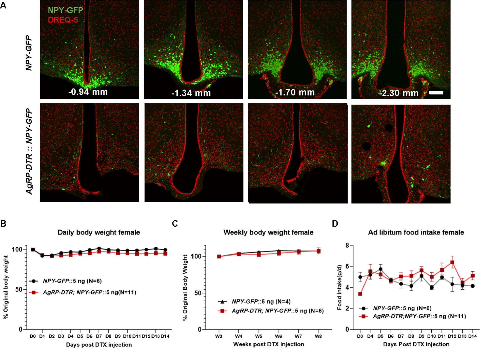
Figure 2. DTX mediated lesion of AgRP neurons in female AgRP-DTR mice.

A, Representative pictures showing GFP (green) and DREQ-5 nucleus staining (red) in NPY-GFP and AgRP-DTR::NPY-GFP female mice with single i.c.v injection of 5 ng DTX injections at a series of sections with the indicated Bregma levels in mm. B-D, Comparison in female body weight during the first 14 days (B) and 8 weeks (C) after toxin injections; and daily food intake (D) at the indicated day after toxin injection. Scale bar = 100 μm. Two-way repeated ANOVA followed by Bonferroni’s multiple comparisons: p = 0.1160 (B); p = 0.6557 (C); p = 0.1171 (D). Data was presented as mean ± SEM.
