Fig. 1.

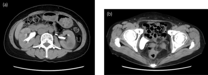
(a) Abdominal contrast CT shows slight hydronephrosis in the right kidney. (b) Contrast accumulation is observed on the right side of the pelvic cavity.

(a) Abdominal contrast CT shows slight hydronephrosis in the right kidney. (b) Contrast accumulation is observed on the right side of the pelvic cavity.