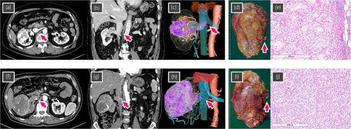
Fig. 1.

(a–e) Case 1 showing right RCC with a level II IVC tumor thrombus (arrow). (a) Axial section of computed tomography (CT). (b) Coronal section of CT. (c) Three‐dimensional reconstructed image from CT. (d), Macroscopic findings of the excised right kidney and IVC tumor thrombus (arrow). Excised weight was 595 g. (e) Microscopic findings of hematoxylin and eosin staining showing clear cell RCC, pT3b, WHO/ISUP grade 3. (f–j) Case 2 showing right RCC with level I IVC tumor thrombus (arrow). (f) Axial section of CT. (g) Coronal section of CT. (h) Three‐dimensional reconstructed image from CT. (i), Macroscopic findings of the excised right kidney and IVC tumor thrombus (arrow). Excised weight was 635 g. (j), Microscopic findings of hematoxylin and eosin staining showing clear cell RCC, pT3b, WHO/ISUP grade 4.
