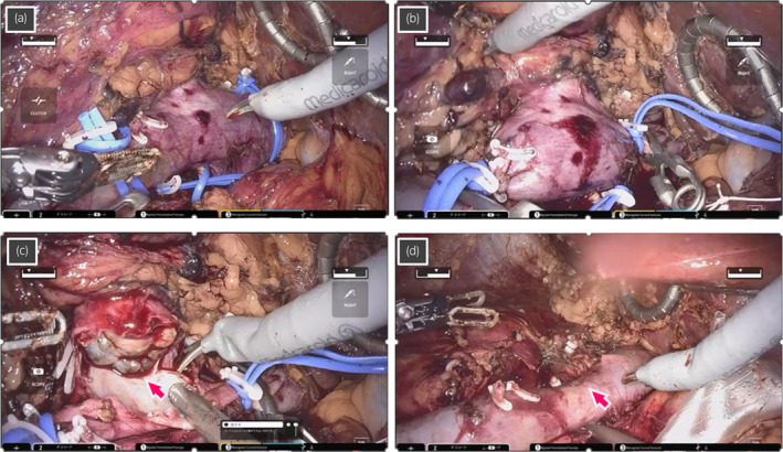
Fig. 2.

Intraoperative images during robot‐assisted RN and IVC tumor thrombectomy for case 2 who had level I IVC tumor thrombus. (a) The left renal vein, caudal IVC, and cephalic IVC are secured by twice‐wrapped vessel loops, and (b) sequentially clamped with the vessel loops by clipping in addition to the use of bulldogs. (c) Tumor thrombus (arrow) is removed from the IVC, and the wall of IVC is cut. (d) The IVC is reconstructed by continuous suture with a 4‐0 polypropylene (arrow), following removal of the tumor thrombus.
