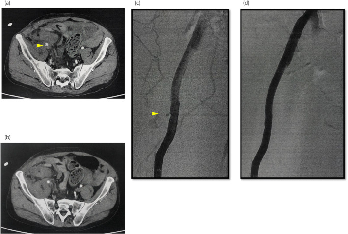
Fig. 2.

Contrast test to identify the bleeding site. (a) CT showing a pseudoaneurysm at the anastomosis of the EIA (b) The contrast material is also exposed to the abdominal cavity (c) Angiography showing the pseudoaneurysm (d) Covered stent placement (thickness, 8 mm; length, 10 cm).
