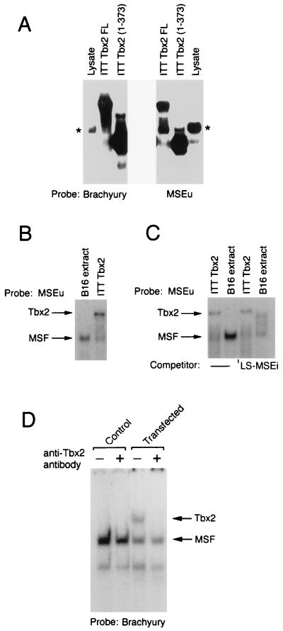
FIG. 3.
Tbx2 can bind the MSEu and MSEi and is distinct from MSF. (A) Band shift assay using a radiolabeled brachyury-binding site or the MSEu as the probe together with either unprogrammed reticulocyte lysate, ITT Tbx2FL, or the C-terminal truncation Tbx2(1-373). Only the bound DNA is shown. The asterisk indicates the position of a complex originating in the unprogrammed reticulocyte lysate and which has a mobility similar to that of MSF. (B) Band shift assay as in panel A, using the MSEu probe and either B16 nuclear extract or ITT Tbx2. The relative positions of the MSF- and Tbx2-containing complexes are indicated. (C) As for panel B but with 50 ng of the indicated LS-MSEi competitor where indicated. (D) Band shift assay using B16 extract derived from untransfected cells (control) or cells transfected with a Tbx2 expression vector, together with a brachyury consensus binding site as a probe. The DNA-binding reaction was performed in the presence or absence of a polyclonal anti-Tbx2 antiserum as indicated.

