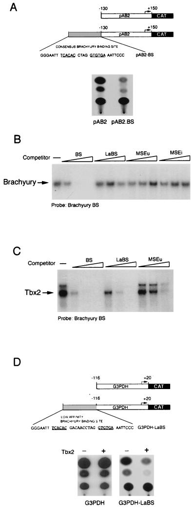
FIG. 5.
Transcriptional repression by Tbx2 in melanocytes. (A) Schematic showing the structure of the herpes simplex virus IE110 promoter (in pAB2) and the consensus brachyury site (BS). (B) Brachyury does not bind the LaBS, the MSEu, or the MSEi, as determined by band shift assay using ITT brachyury together with the consensus brachyury probe and 10, 50, or 250 ng of the indicated consensus BS, LaBS, and MSEu, and MSEi competitors. (C) Tbx2 binds the consensus brachyury site around 5-fold better than the LaBS and around 15-fold better than the MSEu, as determined by band shift assay using ITT Tbx2 together with the consensus brachyury probe and 10, 50, or 250 ng of the consensus (BS) or low-affinity (LaBS) brachyury site or MSEu as the competitor. Only the bound DNA is shown. The full sequences of the BS and LaBS sites are shown in panels A and D, respectively. (D) Transcriptional repression by Tbx2. The indicated G3PDH-CAT or G3PDH.LaBS-CAT reporters were transfected into the melanocyte cell line melan-c either alone or together with a Tbx2 expression vector, and CAT activity was determined 48 h posttransfection.

