
Figure 2.
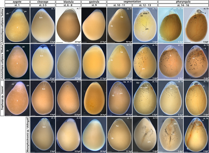
Early embryonic development (zygote to pharyngula). Stage numbering following the staging table of Oreochromis niloticus (Fujimura & Okada, 2007) (see Supporting Information: Table S1 for stage descriptions and associated developmental landmarks). Embryos undergoing somitogenesis are outlined in the “segmentation” stage. Lateral views except for dorsal views in RC st. 12–16. No st. 1 (zygote) image available for RC due to their prolonged courting and breeding behavior. bm, blastomeres; ch, chorion; dpf, days postfertilization; e, eye; em, eye melanophores; ha, head anlagen; hb, hindbrain; l, lens; mhb, midbrain‐hindbrain boundary; ov, otic vesicle; ps, perivitelline space; st, stage; tb, tailbud; y, yolk; ym, yolk melanophores. Scale bar = 1 mm. [Color figure can be viewed at wileyonlinelibrary.com]