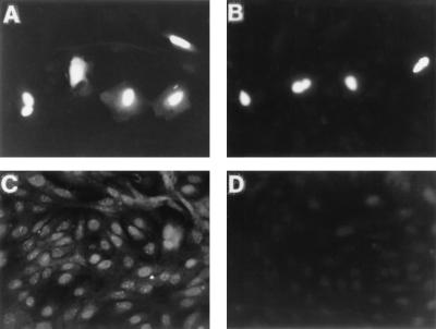
FIG. 3.
Immunocytochemical localization of SNURF in CV-1 cells. CV-1 cells seeded on glass slips on 10-cm plastic plates were transfected by using DOTAP transfection reagent and 1 μg of FLAG-SNURF expression vector as described in Materials and Methods. Cells were fixed in 4% (wt/vol) paraformaldehyde and permeabilized, and the SNURF protein was visualized with rabbit antiserum raised against GST-SNURF (A) or anti-FLAG M2 antibody (B) as described in Materials and Methods. (C) The distribution of endogenous SNURF in CV-1 cells as shown by using anti-SNURF antiserum. (D) Immunofluorescence of CV-1 cells with anti-SNURF antiserum neutralized with purified GST-SNURF fusion protein.

