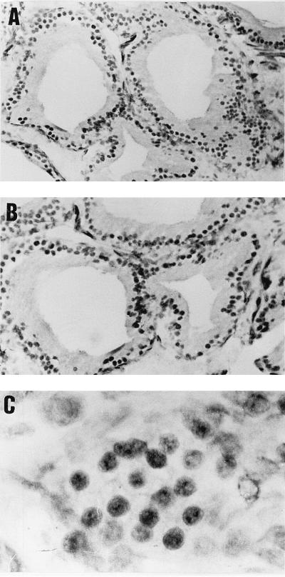
FIG. 4.
Cellular distribution of SNURF and AR in rat prostate. The immunoperoxidase technique was applied to visualize SNURF and AR by using polyclonal antisera as described in Materials and Methods. (A) SNURF is localized in the nuclei of epithelial cells. (B) Immunoreactive AR protein shows the same pattern of distribution as for SNURF. (C) A higher magnification shows granular clusters of SNURF immunoreactivity in epithelial cell nuclei. Magnifications: ×550 (A), ×700 (B), ×2,800 (C).

