
Figure 2.
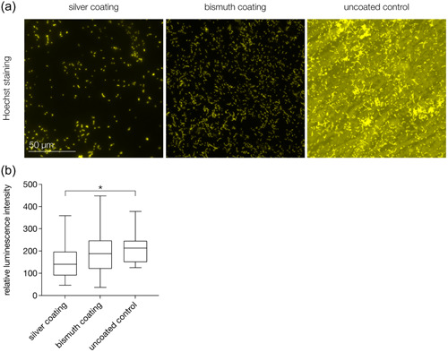
Adhesion of Streptococcus mutans to the investigated silver‐ and bismuth‐coated elastomeric specimens. (a) Hoechst staining shows typical chain formations of S. mutans cells. The surface of the uncoated control shows strong auto fluorescence. There is a tendency for lower bacterial accumulation on silver‐coated specimens. (b) Results of the luminescence assay show significantly lower adhesion of S. mutans to silver‐coated specimens compared to uncoated controls. *p < .05.