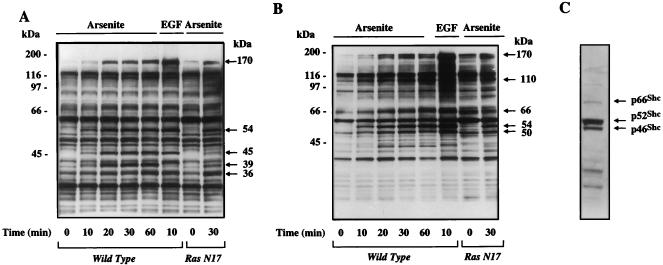
FIG. 2.
Protein tyrosine phosphorylation profile of arsenite-treated PC12 cells. (A) Wild-type and Ras N17 PC12 cells were treated with 400 μM arsenite or 100 ng of EGF per ml for the indicated times. The cell extracts were separated on 10% NuPAGE Bis-Tris gel. Tyrosine-phosphorylated proteins were detected by immunoblotting with the antiphosphotyrosine antibody 4G10. The major tyrosine-phosphorylated bands induced by arsenite or EGF are indicated by arrows. (B) Protein tyrosine phosphorylation profile in arsenite-treated PC12 cells was generated on a 4 to 12% gradient NuPAGE Bis-Tris gel. Tyrosine-phosphorylated proteins were detected by immunoblotting with the antiphosphotyrosine antibody 4G10. Arrows indicate the major tyrosine-phosphorylated bands induced by arsenite or EGF. (C) Shc isoforms comigrate with tyrosine-phosphorylated proteins induced by arsenite or EGF. The blot shown in panel B was stripped and reprobed with an Shc-specific rabbit polyclonal antibody. Shown is a single lane from this blot (wild-type, arsenite-treated cells), as all lanes showed identical patterns.

